The Heart
What is it?
Your heart is a fist-sized organ that pumps blood throughout your body. It’s your circulatory system’s main organ. Muscle and tissue make up this powerhouse organ.
How does it function?
-Your heart’s main function is to circulate blood throughout your body, during circulation the blood brings oxygen and nutrients to your cells. It also takes away carbon dioxide and other waste so other organs can dispose of them.
Other functions of the heart include:
Maintaining the heart rate, both speed and rhythm
Regulating blood pressure
Your heart interacts with these systems to control several different bodily functions:
Nervous system: Your nervous system helps control your heart rate. It sends signals that tell your heart to beat slower or faster depending on the level of stress you are experiencing
Endocrine system: Your endocrine system sends out hormones. Said hormones tell your blood vessels to constrict or relax, which affects your blood pressure. Hormones from your thyroid gland can also tell your heart to beat faster or slower.
Your heart anatomy includes:
Walls
Chambers
Valves
Blood vessels
An electrical conduction system
Anatomy of the Heart
Your heart walls are the muscles that contract and relax to send blood throughout your body. A layer of muscular tissue called the septum divides your heart walls into the left and right sides.
Your heart walls have three layers:
Endocardium: Inner layer.
Myocardium: Muscular middle layer
Epicardium: Protective outer layer
The epicardium is one layer of your pericardium. The pericardium is a protective sac that covers your entire heart. It produces a fluid to lubricate your heart and keep it from rubbing against other organs.

Heart Walls
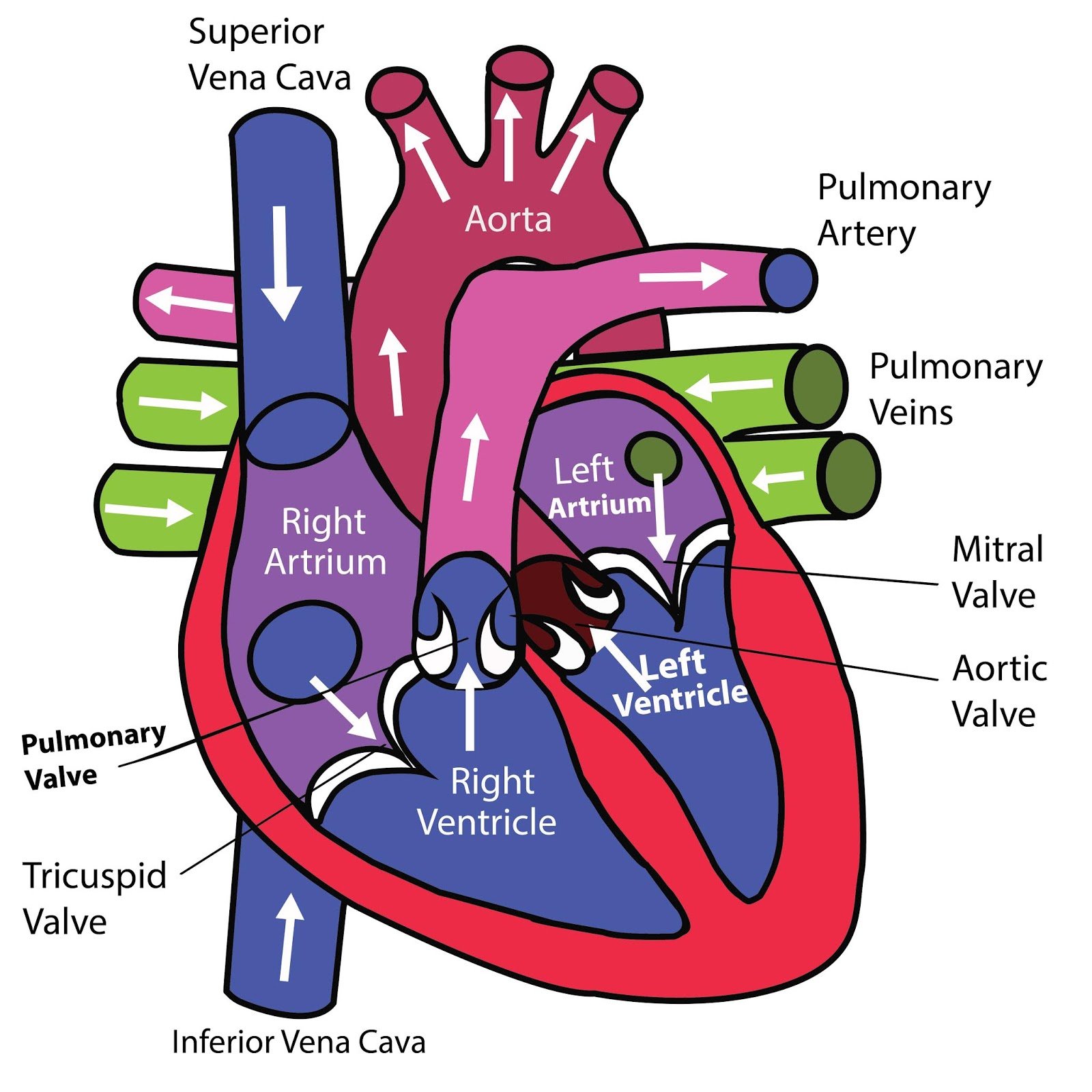
Your heart has four chambers. There are two chambers on the top (atrium) and two on the bottom (ventricles), one on each side of your heart.
Right atrium: Two large veins deliver oxygen-poor blood to your right atrium. The superior vena cava carries blood from your upper body. The inferior vena cava brings blood from your lower body. Then the right atrium pumps blood to your right ventricle.
Right ventricle: The lower right chamber pumps oxygen-poor blood into your lungs through the pulmonary artery. The lungs reload blood with oxygen.
Left atrium: After the lungs fill your blood with oxygen, the pulmonary veins carry the blood to the left atrium. This upper chamber pumps blood to your left ventricle.
Left ventricle: The left ventricle is slightly larger than the right. It pumps oxygen-rich blood into the rest of your body.
Heart Valves

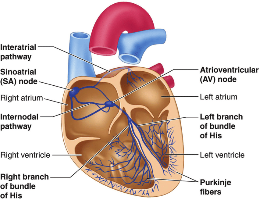
Electrical Conduction System
Your hearts conduction system like the electrical wiring of a building. It controls the rhythm and pace of your heartbeat. Signals start at the top of your heart and move down to the bottom. Your conduction system includes:
Sinoatrial (SA) node: Sends the signals that make your heart beat.
Atrioventricular (AV) node: Carries electrical signals from your heart’s upper chambers to its lower ones.
Left branch of bundle of His: Sends electric impulses to your left ventricle.
Right branch of bundle of His: Sends electric impulses to your right ventricle.
Bundle of His: Sends impulses from your AV node to the Purkinje fibers.
Purkinje fibers: Make your heart ventricles contract and pump out blood.
Your heart is in the front of your chest. It sits slightly behind and to the left of your sternum, which is in the middle of your chest.
Your heart is slightly on the left side of your body. It sits between your right and left lungs. The left lung is slightly smaller to make room for the heart in your left chest. Your ribcage protects your heart.
Common Conditions of the Heart
Arrhythmia: A heartbeat that’s too fast, too slow or beats with an irregular rhythm.
Cardiomyopathy: Unusual thickening, enlargement or stiffening of your heart muscle.
Congestive Heart: Your heart is too stiff or too weak to properly pump blood throughout your body.
Coronary Artery disease: Plaque buildup that leads to narrow coronary arteries.
Diabetes: Your blood sugar is higher than it should be.
Heart Attack: A sudden coronary artery blockage that cuts off oxygen to part of your heart muscle.
Heart Valve Disease: A valve in your heart isn’t working right.
High Blood Pressure: Your blood is pushing too hard against your artery walls.
High Cholesterol: Your blood has too many fats in it.
Pericarditis: Inflammation in your heart’s lining (pericardium).

Your heart valves open and close to allow blood to flow through. They also keep your blood from moving in the wrong direction.
Atrioventricular valves
The atrioventricular (AV) valves open between your upper and lower heart chambers. They include:
Tricuspid valve: Valve between your right atrium and right ventricle.
Mitral valve: Valve between your left atrium and left ventricle.
Semilunar valves: Semilunar (SL) valves open when blood flows out of your ventricles.
Aortic Valve: Opens when blood flows out of your left ventricle to your aorta (artery that carries oxygen-rich blood to your body).
Pulmonary Valve: Opens when blood flows from your right ventricle to your pulmonary arteries (the only arteries that carry oxygen-poor blood to your lungs).
Where is your heart located?
Common Signs of heart condition
Chest pain
Heart palpitations
Dizziness
Shortness of breath
Fatigue
Swelling in your lower body
The Brain
What is the brain? The brain is a complex organ that has control over thought, memory, emotion, touch, motor skills, vision, breathing, temperature, hunger and every process that regulates our body. The brain and spinal cord make up the central nervous system, or CNS.
What is it made up of? The brain is about 60% fat. The remaining 40% is a combination of water, protein, carbohydrates and salts. The brain itself is not a muscle. It contains blood vessels and nerves, including neurons and glial cells. It is grey and white matter.
What is grey and white matter? Gray and white matter are different regions of the central nervous system. In the brain, gray matter refers to the darker, outer portion (the Dura), while white matter describes the lighter, inner section underneath.
In the spinal cord, this order is reversed: The white matter is on the outside, and the gray matter sits within. Gray matter is primarily composed of neuron somas (the round central cell bodies), white matter is mostly made of axons (the long stems that connects neurons together) wrapped in myelin (a protective sheath). The different make up of neuron parts is why the two appear as separate shades on certain scans.
How does the brain function?
The brain sends and receives chemical and electrical signals throughout the body. Different signals control different processes, your brain interprets each. Some messages are kept within the brain, while others are sent through the spine and across the body’s vast network of nerves to distant extremities. The central nervous system relies on billions of neurons (nerve cells) to send and receive all the messages the body makes through throughout the day
Anatomy
Cerebrum - The cerebrum, the front of the brain, is the largest part of the brain, the cerebrum initiates and coordinates movement and regulates temperature. Other areas of the cerebrum enable speech, judgment, thinking and reasoning, problem-solving, emotions and learning. Other functions relate to vision, hearing, touch and other senses.
Cerebral Cortex -The cortex has a large surface area due to its folds, and comprises about half of the brain’s weight. The cerebral cortex is divided into two halves. It is covered with ridges (gyri) and folds (sulci). The two halves join at a large, deep sulcus (the interhemispheric fissure, AKA the medial longitudinal fissure) that runs from the front of the head to the back. The right hemisphere controls the left side of the body, and the left half controls the right side of the body. The two halves communicate with one another through a large, C-shaped structure of white matter and nerve pathways called the corpus callosum. The corpus callosum is in the center of the cerebrum.
Brainstem The brainstem (middle of brain) connects the cerebrum with the spinal cord. The brainstem includes the midbrain, the pons and the medulla.
Midbrain. The midbrain (mesencephalon) is a very complex structure with a range of different neuron clusters (nuclei and colliculi), neural pathways and other structures. These features facilitate various functions, from hearing and movement to calculating responses and environmental changes. The midbrain also contains the substantia nigra, an area affected by Parkinson’s disease that is rich in dopamine neurons and part of the basal ganglia, which enables movement and coordination.
The Brain Coverings
Three layers of protective covering called meninges surround the brain and the spinal cord.
The outermost layer, the dura mater, is thick and tough. It includes two layers: The periosteal layer of the dura mater lines the inner dome of the skull (cranium) and the meningeal layer is below that. Spaces between the layers allow for the passage of veins and arteries that supply blood flow to the brain.
The arachnoid mater is a thin, weblike layer of connective tissue that does not contain nerves or blood vessels. Below the arachnoid mater is the cerebrospinal fluid, or CSF. This fluid cushions the entire central nervous system (brain and spinal cord) and continually circulates around these structures to remove impurities.
The pia mater is a thin membrane that hugs the surface of the brain and follows its contours. The pia mater is rich with veins and arteries.

Pons. The pons is the origin for four of the 12 cranial nerves, which enable a range of activities such as tear production, chewing, blinking, focusing vision, balance, hearing and facial expression. Named for the Latin word for “bridge”, the pons is the connection between the midbrain and the medulla.
Medulla. At the bottom of the brainstem, the medulla is where the brain meets the spinal cord. The medulla is essential to survival. Functions of the medulla regulate many bodily activities, including heart rhythm, breathing, blood flow, and oxygen and carbon dioxide levels. The medulla facilitates reflexive activities such as sneezing, vomiting, coughing and swallowing.
The spinal cord extends from the bottom of the medulla and through a large opening in the bottom of the skull. Supported by the vertebrae, the spinal cord carries messages to and from the brain and the rest of the body.
Cerebellum - The cerebellum (“little brain”) is a fist-sized portion of the brain located at the back of the head, below the temporal and occipital lobes and above the brainstem. Like the cerebral cortex, it has two hemispheres. The outer portion contains neurons, and the inner area communicates with the cerebral cortex. Its function is to coordinate voluntary muscle movements and to maintain posture, balance and equilibrium. Current studies are exploring the cerebellum’s roles in thought, emotions and social behavior, as well as its possible involvement in addiction, autism and schizophrenia.
Lobes of the Brain
Frontal lobe. The largest lobe of the brain, located in the front of the head, the frontal lobe is involved in personality characteristics, decision-making and movement. Recognition of smell usually involves parts of the frontal lobe. The frontal lobe contains Broca’s area, which is associated with speech ability.
Parietal lobe. The middle part of the brain, the parietal lobe helps a person identify objects and understand spatial relationships (where one’s body is compared with objects around the person). The parietal lobe is also involved in interpreting pain and touch in the body. The parietal lobe houses Wernicke’s area, which helps the brain understand spoken language.
Occipital lobe. The occipital lobe is the back part of the brain that is involved with vision.
Temporal lobe. The sides of the brain, temporal lobes are involved in short-term memory, speech, musical rhythm and some degree of smell recognition.
Other Structures of the Brain
Pituitary Gland
Sometimes called the “master gland,” the pituitary gland is a pea-sized structure found deep in the brain behind the bridge of the nose. The pituitary gland governs the function of other glands in the body, regulating the flow of hormones from the thyroid, adrenals, ovaries and testicles. It receives chemical signals from the hypothalamus through its stalk and blood supply.
Hypothalamus
The hypothalamus is located above the pituitary gland and sends it chemical messages that control its function. It regulates body temperature, synchronizes sleep patterns, controls hunger and thirst and also plays a role in some aspects of memory and emotion.
Amygdala
Small, almond-shaped structures, an amygdala is located under each half (hemisphere) of the brain. Included in the limbic system, the amygdalae regulate emotion and memory and are associated with the brain’s reward system, stress, and the “fight or flight” response when someone perceives a threat.

Two sets of blood vessels supply blood and oxygen to the brain: the vertebral arteries and the carotid arteries.
The external carotid arteries ascend the sides of your neck, are where you can feel your pulse when you touch the area with your fingertips. The internal carotid arteries branch into the skull and circulate blood to the front part of the brain.
The vertebral arteries follow the spinal column into the skull, where they join together at the brainstem and form the basilar artery, which supplies blood to the rear portions of the brain.
The circle of Willis, a loop of blood vessels near the bottom of the brain that connects major arteries, circulates blood from the front of the brain to the back and helps the arterial systems communicate with one another.
Hippocampus
A curved seahorse-shaped organ on the underside of each temporal lobe, the hippocampus is part of a larger structure called the hippocampal formation. It supports memory, learning, navigation and perception of space. It receives information from the cerebral cortex and may play a role in Alzheimer’s disease.
Pineal Gland
The pineal gland is located deep in the brain and attached by a stalk to the top of the third ventricle. The pineal gland responds to light and dark and secretes melatonin, which regulates circadian rhythms and the sleep-wake cycle.
Ventricles and Cerebrospinal Fluid
Deep in the brain are four open areas with passageways between them. They also open into the central spinal canal and the area beneath arachnoid layer of the meninges.
The ventricles manufacture cerebrospinal fluid, or CSF, a watery fluid that circulates in and around the ventricles and the spinal cord, and between the meninges. CSF surrounds and cushions the spinal cord and brain, washes out waste and impurities, and delivers nutrients.
Blood Supply to the Brain

What are they?
The kidneys are two bean-shaped organs that filter your blood and are part of your urinary system. Your kidneys filter roughly 200 quarts of fluid every day. During the filtration process, the kidneys remove waste, which leaves your body as urine. Most people pee about two quarts daily. Your body re-uses the other 198 quarts of fluid. Your kidneys also help balance your body’s fluids and electrolytes. Electrolytes are essential minerals that include sodium and potassium.
The Kidneys

How do the kidneys function?
Blood flows into your kidneys through a large blood vessel called the renal artery.
Tiny blood vessels in your kidney filter the blood.
The filtered blood returns to your bloodstream through a large blood vessel called the renal vein.
Pee travels through tubes of muscle called ureters to your bladder.
Your bladder stores pee until you release it through urination (peeing).
The kidneys have many important functions, they clean toxins and waste out of your blood. Common waste products include but are not limited to nitrogen waste (urea), muscle waste (creatinine) and acids. They help your body remove these substances. The kidneys filter about half a cup of blood every minute.
Other functions of the kidney include:
Control the acid-base balance, pH balance, of your blood.
Make sugar (glucose) if your blood doesn’t have enough sugar.
Make a protein called renin that increases blood pressure.
Produce the hormones calcitriol and erythropoietin. (Calcitriol is a form of vitamin D that helps your body absorb calcium. Erythropoietin helps your body make red blood cells.)
An adrenal gland sits on top of each kidney. It produces hormones, including cortisol, which helps your body respond to stress.
How does blood filtration work? Each kidney contains more than a million filtering units called nephrons. Each nephron is made up of:
Glomeruli: Glomeruli are groups of tiny blood vessels that perform the first stage of filtering your blood. They then pass filtered substances to the renal tubules. The name for this process is glomerular filtration.
Renal tubules: These tiny tubes reabsorb and return water, nutrients and minerals your body needs. The tubules remove waste, through a process called diffusion. Your body sends the remaining waste through your kidneys’ collecting chambers. In its final act =, it leaves your body as pee.
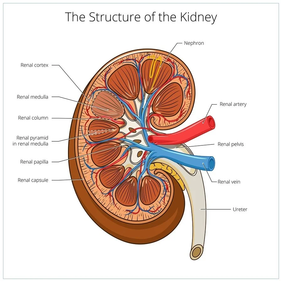
Anatomy of the Kidneys
Your kidneys sit below your ribcage and behind your stomach. Typically, one kidney sits on either side of your spine. Your kidneys reside between your intestines and diaphragm. A ureter connects each kidney to your bladder. The kidney is roughly 4-5 inches long
Kidney capsule
The kidney capsule consists of three layers of connective tissue or fat that cover your kidneys. It protects your kidneys from injury, increases their stability and connects your kidneys to surrounding tissues.
Renal artery
The renal artery is a large blood vessel that controls blood flow into your kidneys. For most people at rest, the renal kidneys pump a little over 5 cups of blood to your kidneys each minute.
Renal cortex
The outer layer of your kidney, where the nephrons begin. The renal cortex also creates the hormone erythropoietin (EPO), which helps make red blood cells in your bone marrow.
Signs of complications within the kidneys
Most kidney problems don’t have signs in their early stages. As kidney damage progresses, you may notice:
Cramping muscles: Electrolyte imbalances cause your muscles to stiffen.
Dark urine or urine with blood in it: Damage to your kidneys’ filters lets blood cells leak into your urine.
Foamy urine: Bubbles in your pee can signal excess protein.
Itchy, dry skin: An imbalance of minerals and nutrients in your blood leads to itchy skin.
More frequent urination: Problems filtering waste cause you to pee more often.
Puffy eyes or swollen ankles and feet: Reduced kidney function can cause your body to hold onto protein and sodium, resulting in swelling.
Sleep problems, fatigue and lack of appetite: If toxins build up in your blood, your sleep, appetite and energy levels may be off.
Renal medulla
The renal medulla is the inner part of your kidney. It contains most of the nephrons with their glomeruli and renal tubules. The renal tubules carry urine to the renal pelvis.
Renal papilla
These pyramid-shaped structures transfer urine to the ureters. Dehydration and certain medications, especially nonsteroidal anti-inflammatory drugs (NSAIDs) can cause damage your renal papilla.
Renal pelvis
This funnel-shaped structure collects urine and passes it down two ureters. Urine travels from the ureters to the bladder, where it’s stored.
Renal vein
This vein is the main blood vessel that carries filtered blood out of your kidneys and back to your heart. Each of your kidneys has a renal vein.
How to keep your kidney’s healthy
Avoid smoking and any tobacco products
Decrease the use of excessive salt, salt can affect the balance of minerals in your blood.
Drink water more than you drink sugary or processed drinks
Decrease the amount of time you remain sedentary, exercise will help to regulate your blood pressure
Avoid use of NSAIDs. NSAIDs can cause kidney damage if you take them too much.
Limit weight fluctuation, maintain a healthy weight
Monitor your blood pressure, When the blood pressure is unstable there could be something in the kidneys
If you are a diabetic, monitor your blood sugar anything out of the normal should be checked by your doctor

The Lungs
What are the lungs? Your lungs make up a large part of your respiratory system, which is the network of organs and tissues that allow you to breathe. You have two lungs, one on each side of your chest, which is also called the thorax. Your thorax is the area of your body between your neck and your abdomen.
Right lung - The lung on your right side is divided into three lobes: the superior, the middle and the inferior. It’s shorter than your left lung, but also wider than your left lung. Both of your lungs are covered with a protective sheath called pleural tissue.
Left lung - Your left lung has two lobes: the superior and the inferior. Your left lung is smaller than the right because your heart sits where a middle lobe would be. Your left lung has two parts that your right lung doesn’t have: the cardiac notch (where your heart fits) and the lingula, an extension of the superior lobe
How do the lungs function? Your lungs provide an exchange of oxygen and gas, You breathe in oxygen while breathing out other gases your body produces, like carbon dioxide. This process should take place about 12 to 20 times per minute. When you inhale through your nose or mouth, air travels down your back of your throat (pharynx), passes through your voice box (larynx) and into your windpipe (trachea). Your trachea is divided into two air passages, the bronchial tubes. One bronchial tube leads to your left lung, the other to your right lung.
This function allows your lungs to perform at an optimal state, your airways need to be open when you inhale and when you exhale. They also need to be free from inflammation and abnormal amounts of mucus. Your bronchial tubes lead to smaller air passages called bronchi then into bronchioles. The bronchioles end in tiny air sacs called alveoli, where oxygen is transferred from the inhaled air to your blood. Alveoli look like clusters of tiny grapes.
After absorbing oxygen, the blood leaves your lungs and is carried to your heart. From there, it’s pumped through your body to provide oxygen to the cells of your tissues and organs. When cells use oxygen, they produce carbon dioxide which is transferred to your blood. Your bloodstream carries the carbon dioxide back to your lungs. When you exhale, you remove the carbon dioxide.
Your respiratory system prevents harmful substances from entering your lungs by using:
Small hairs in your nose. They act as an air-cleaning system and help filter out large particles. This is part of the formation of boogers.
Mucus is produced in your trachea and bronchial tubes to keep air passages moist and help catch dust, bacteria and other substances.
The sweeping motion of small hairs in your respiratory tract called cilia to keep air passages clean.
Anatomy
Your lungs are located in your chest. Your thoracic cavity (chest cavity) is the name of the space that contains your lungs and other organs. Your lungs rest on a muscle called your diaphragm.
A healthy lung is pinkish gray in color. Damaged lungs are darker gray and can have black spots in them, those comparison photos of a smoker’s lung vs a healthy lung are a good reference.
Interesting facts about your lungs
You can have lobes of your lung removed and live. You can even live with only one lung.
Lungs are the only organs in your body that will float.
Exercise can help you increase your lung capacity.
A typical adult has 300 million to 500 million alveoli.
Signs of trouble in the lungs
Common signs and symptoms of lung conditions include:
Shortness of breath
Chest pain
Coughs, chronically, and coughing up blood
Fatigue
Wheezing
Swelling in your ankles and feet.
What is the Liver? The liver is an essential organ, performing hundreds of functions necessary to sustain life. It's also a gland because it makes proteins and hormones that other parts of the body need. Under normal conditions, the liver is located on the right side of the body, under the ribs. In a condition called situs inversus, the liver is located on the left side.
Anatomy - The liver has two main parts: the larger right lobe and the smaller left lobe.
The lobes contain many blood vessels. Blood travels through the liver, the liver filters, or cleans, the blood, removing toxins and waste that eventually leave the body through urine and feces.
The lobes also contain thousands of lobules (small lobes). These lobules connect with many bile ducts, tubes that transport bile from the liver to the small intestine.
The Liver

What are the symptoms of liver problems?
When a person has a liver problem, one of the most common symptoms is Jaundice.
With jaundice, the skin and whites of the eyes turn yellow because of too much bilirubin in the blood. Bilirubin is a yellow waste product the liver gets rid of when it breaks down red blood cells. Higher levels of bilirubin indicate a possible problem in the liver.
Other symptoms of liver problems may include:
Build-up of fluid in the belly area
Easy bruising
Itchy skin
Low blood sugar
Pain in the abdomen
Swelling in the ankles
Tremors
Weakness, loss of balance or constant fatigue.
How does the Liver Function? The liver has hundreds of jobs. Some of the most vital are:
· Cleans toxins out of the blood.
· Gets rid of old red blood cells.
· Makes bile, a fluid that helps the body digest food.
· Metabolizes proteins, carbohydrates and fats so your body can use them.
· Produces substances to help blood clot.
· Regulates the amount of blood in the body.
· Stores glycogen and vitamins to be used by the body later.
What conditions and disorders affect the liver?
Many conditions can affect the liver. Among the most common are:
Diseases occurring when a person consumes toxins in excess amounts, such alcohol related liver disease and fatty liver disease
Hereditary diseases like hemochromatosis and Wilson Disease
Liver Cancer
Problems when the immune system attacks the liver, such as autoimmune hepatitis,
Viral infections, such as Hepatitis A,B and C
Many of these conditions can lead to cirrhosis.
Sometimes, damaged liver tissue can regenerate or grow back. Other times, liver disease can cause serious symptoms and even be life-threatening.
How can I keep my liver healthy?
To keep your liver healthy and functioning well, try to follow these tips:
Avoid toxins, such as chemicals, smoking and illegal drugs.
Don’t share needles, razors, toothbrushes or any other personal items, which can spread viruses.
Drink alcohol only in moderation.
Follow healthcare professionals’ instructions about medications, especially warnings against mixing medications and alcohol.
Maintain a healthy weight, including eating a nutritious diet and exercising regularly.
Practice safe sex to avoid hepatitis infection.
Talk to your healthcare provider about vaccinations against hepatitis.
Wash your hands frequently.